


- CaBLAM makes neurons glow without external light at single-cell resolution.
- Bioluminescence eliminates phototoxicity, photobleaching, and invasive fiber optics.
- Self-lit neurons produce signal against a dark background, improving clarity.
Neuroscientists at Brown University just figured out how to watch individual brain cells fire in real time - without shining any light into the brain at all.
The trick: engineer neurons to glow like fireflies.
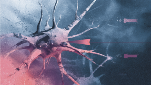
The Brown team created a bioluminescent probe called CaBLAM that finally makes brain cells bright enough to track single neurons without external illumination.

Key figure
1st
Bioluminescent probe to achieve single-cell resolution in living brain tissue
Why Standard Brain Imaging Creates Problems
For years, neuroscientists have relied on fluorescent proteins to monitor brain activity. The approach works: shine laser light at engineered cells, and they emit a different color back when calcium flows during neural firing.
But that external light creates three serious issues.
First, bombarding tissue with intense beams damages cells over time, a condition called phototoxicity.
Second, prolonged illumination causes photobleaching, where the fluorescent molecules degrade and stop emitting adequate signal.
Third, delivering laser light deep into brain tissue requires invasive hardware like fiber optics threaded through living neural circuits.
Christopher Moore, who leads Brown's Bioluminescence Hub, points out another challenge: brain tissue naturally glows faintly when hit by external light, creating background noise that obscures the signals researchers want to measure.
The Firefly Solution
Bioluminescence sidesteps all these problems by making neurons produce their own light through a chemical reaction. An enzyme breaks down a small molecule, releasing photons without any external beam required.
The concept isn't new. Scientists have discussed bioluminescent brain imaging for decades. But nobody could make the glow bright enough to capture detailed activity from individual cells.
CaBLAM changes that equation, as explained in a Nature open-access paper in December 2025.
The probe responds to calcium, which floods into neurons when they fire. When calcium concentration rises, the bioluminescent signal intensifies.
What is calcium imaging?
Neurons release a burst of calcium ions every time they fire. By engineering brain cells to change their light output in response to calcium, scientists can watch which neurons activate and when. Traditional methods require external lasers to trigger this glow; bioluminescent probes like CaBLAM generate their own light instead.
As Shaner puts it, the neurons become their own headlights. Researchers only need to capture the light coming out, which penetrates scattered brain tissue far more clearly than reflected external beams.
What makes this significant is the signal-to-noise advantage. Since brain tissue doesn't naturally produce bioluminescence, active neurons glow against a completely dark background.
No scattered laser light. No ambient tissue fluorescence. Just the cells that are firing.
...with bioluminescence, the brain cells act like their own headlights
Nathan Shaner, University of California at San Diego
What Single-Cell Resolution Unlocks
Moore emphasizes that CaBLAM represents the first time bioluminescent probes have achieved single-cell resolution.
Scientists can now record independent neural activation patterns almost like running a highly sensitive movie camera inside living brain circuits.
This opens direct pathways to experiments that were previously too risky or technically impossible. Long-term imaging studies become feasible without cumulative light damage.
More On Brain Cells
A Brain Cell That Always Knows Which Way You're Facing
Scientists found a neuron that tracks your direction whether you're sitting still or running–and losing it may explain Alzheimer's disorientation.
→Deep brain structures become accessible without threading hardware through fragile tissue. Chronic recordings can run indefinitely without photobleaching ending the experiment.
The shift from fluorescence to bioluminescence mirrors a broader pattern in neuroscience: the most powerful tools often emerge not from incremental improvements to existing methods, but from reconsidering fundamental assumptions about how measurements should work.
By eliminating external light entirely, CaBLAM doesn't just reduce problems with conventional imaging. It creates an entirely different optical environment where brain cells become visible in ways that scattered laser beams could never achieve.

Fact Check: Claim-by-Claim Verification Verified
The recap accurately represents the Brown University press release and the underlying Nature Methods paper on CaBLAM, with correct details on researchers, probe function, advantages over fluorescence, and bioluminescent imaging capabilities.
Commentary
- Article states December 2025 Nature paper, but preprint versions appeared earlier (2023); final publication timing is consistent with recent release.
- Dramatic phrasing like "glow like fireflies" and "crack a long-standing puzzle" is acceptable for popular science recap.
Sources used for verification
Academic/Peer-reviewed:
- CaBLAM: a high-contrast bioluminescent Ca2+ indicator derived... - PubMed
- CaBLAM! A high-contrast bioluminescent Ca2+ indicator derived... - PMC
- Nature Methods publication on CaBLAM - Nature
Other reliable sources:
- Is bioluminescence the key to safe, effective brain imaging? - Brown.edu
- Christopher Moore - Carney Institute - Brown.edu
- About Us - The Bioluminescence Hub - Bioluminescencehub.org
Fact-checked by Perplexity Sonar Pro on 2025-12-29


