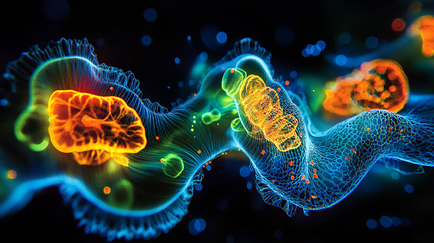

- Fluorophores absorb light at one wavelength and emit at another, enabling targeted molecular imaging.
- Two Nobel Prizes in Chemistry recognized fluorophore imaging advances in 2008 and 2014.
- Super-resolution methods now achieve detail below 50 nanometers, ten times sharper than conventional microscopy.
Fluorophore imaging techniques are methods that use fluorescent molecules to make specific biological structures visible under a microscope, allowing researchers to track proteins, cells, and molecular interactions in living tissue.
Why It Matters
Before fluorophores, biologists worked largely in the dark. They could see cell shapes and boundaries, but the proteins driving disease, signaling between neurons, and building tissue remained invisible. Fluorophore imaging changed that by letting researchers tag individual molecules with glowing labels and watch them move in real time.
Key figure
2
Nobel Prizes in Chemistry awarded for fluorophore imaging breakthroughs (2008 and 2014)
The impact has been enormous. Fluorophore imaging techniques underpin modern cell biology, neuroscience, and cancer research. Two Nobel Prizes in Chemistry, in 2008 and 2014, recognized scientists who advanced the field. The first honored the discovery and development of green fluorescent protein (GFP). The second honored super-resolution fluorescence microscopy, which broke the optical diffraction limit.
Today, fluorophore-based methods are standard tools in labs worldwide. They allow researchers to observe individual neurons firing, track how cancer cells migrate through tissue, and measure calcium ion concentrations inside living cells at high resolution.
How Fluorophore Imaging Works
A fluorophore is a molecule that absorbs light at one wavelength and emits it at a longer wavelength. This shift, called the Stokes shift after Irish physicist George Gabriel Stokes who first described fluorescence in 1852, is what makes the technique possible. By filtering out the excitation light and collecting only the emitted light, a microscope can isolate the signal from tagged molecules against a dark background.
Researchers attach fluorophores to specific targets, such as antibodies that bind a particular protein or genetic constructs that make cells produce fluorescent proteins. When illuminated with the correct wavelength, only the tagged structures glow. Everything else stays dark.
Key figure
~1 μs
Typical delay between photon absorption and fluorescence emission
The process happens fast. Fluorescence emission occurs within roughly one microsecond of excitation. A dichroic mirror inside the microscope reflects the shorter excitation wavelength toward the specimen while letting the longer emission wavelength pass through to a camera or detector. This optical separation is the core engineering principle behind every fluorescence microscope.
Major Fluorophore Imaging Techniques
Several distinct methods have emerged from fluorophore technology, each suited to different biological questions.
Widefield fluorescence microscopy illuminates the entire sample at once. It is fast and simple but captures out-of-focus light, which blurs the image. It works best for thin specimens or surface-level observations.
Confocal microscopy uses a pinhole to reject out-of-focus light, producing sharper optical sections. Researchers can stack these sections to build three-dimensional images of thick tissue. The tradeoff is slower imaging speed.
Two-photon microscopy uses infrared light that penetrates deeper into tissue. Two lower-energy photons combine to excite the fluorophore, limiting fluorescence to a tiny focal volume. Neuroscientists use this method to image neurons deep inside living brains.
Super-resolution microscopy (including STED, PALM, and STORM) pushes past the diffraction limit of visible light. Stefan Hell developed STED at the Max Planck Institute in the 1990s. Eric Betzig and William Moerner independently developed single-molecule methods. These approaches achieve resolution below 50 nanometers, roughly ten times sharper than conventional light microscopy.
Fluorescence resonance energy transfer (FRET) measures the distance between two fluorophores attached to different molecules. When they are close enough (typically under 10 nanometers), energy transfers from one to the other. This reveals whether two proteins interact inside a cell.
Key Context
Japanese chemist Osamu Shimomura isolated green fluorescent protein from the jellyfish Aequorea victoria in 1962. For three decades, GFP remained a curiosity. Then in 1994, Columbia University biologist Martin Chalfie showed that the gene for GFP could be inserted into other organisms, making their cells glow green without any external dye. Roger Tsien at UC San Diego later engineered GFP variants that fluoresce in blue, cyan, and yellow, giving researchers a palette of colors for labeling multiple targets simultaneously.
The 1930s marked the first use of fluorescent dyes (fluorochromes) in biological research for staining tissues and pathogens. By the 2020s, researchers had access to hundreds of synthetic fluorophores spanning the full visible spectrum and into the near-infrared, each optimized for brightness, photostability, or compatibility with specific imaging hardware.
FAQ
What is the difference between a fluorophore and a fluorochrome?
The terms are often used interchangeably, but fluorophore technically refers to the part of a molecule responsible for fluorescence, while fluorochrome refers to the complete fluorescent dye molecule. In practice, most biologists treat them as synonyms.
Can fluorophore imaging be used on living cells?
Yes. Many fluorophores, including genetically encoded fluorescent proteins like GFP, work in living cells without killing them. Live-cell imaging lets researchers watch processes such as protein trafficking, cell division, and signal transduction in real time.
Why do fluorophores photobleach?
Prolonged exposure to excitation light causes irreversible chemical changes in fluorophore molecules, destroying their ability to fluoresce. This effect, called photobleaching, limits how long a sample can be imaged. Researchers manage it by reducing light intensity, using more photostable dyes, or capturing images quickly.
How did super-resolution microscopy overcome the diffraction limit?
Conventional light microscopy cannot resolve structures smaller than about 200 nanometers. Super-resolution methods such as STED use a second laser to selectively switch off fluorophores around a target point, while PALM and STORM image single molecules one at a time and reconstruct their positions computationally. Both approaches achieve resolution below 50 nanometers.
Related Reading
Sources
- Primary Sources:
- Introduction to Fluorescence Microscopy (Nikon MicroscopyU)
- Fluorescence Microscopy: A Concise Guide to Current Imaging Methods (Fiolka et al., PMC, 2013)
- Fluorescence Microscopy: Hardware, Handling, and Fluorophore Considerations (PMC, 2022)
- Additional Context:
- Nobel Prize in Chemistry 2008 (NobelPrize.org)
- Nobel Prize in Chemistry 2014 (NobelPrize.org)
- Fluorescent Proteins: From Beginnings to Nobel Prize (Leica Microsystems)

Fact Check: Claim-by-Claim Verification Verified
All major claims verified against authoritative sources including Nikon MicroscopyU, NobelPrize.org, and peer-reviewed PMC articles. No inaccuracies found.
Sources used for verification
- Introduction to Fluorescence Microscopy - microscopyu.com
- Nobel Prize Chemistry 2008 - nobelprize.org
- Nobel Prize Chemistry 2014 - nobelprize.org
- Fluorescence Microscopy: Concise Guide - pmc.ncbi.nlm.nih.gov
- Fluorescent Proteins History - leica-microsystems.com